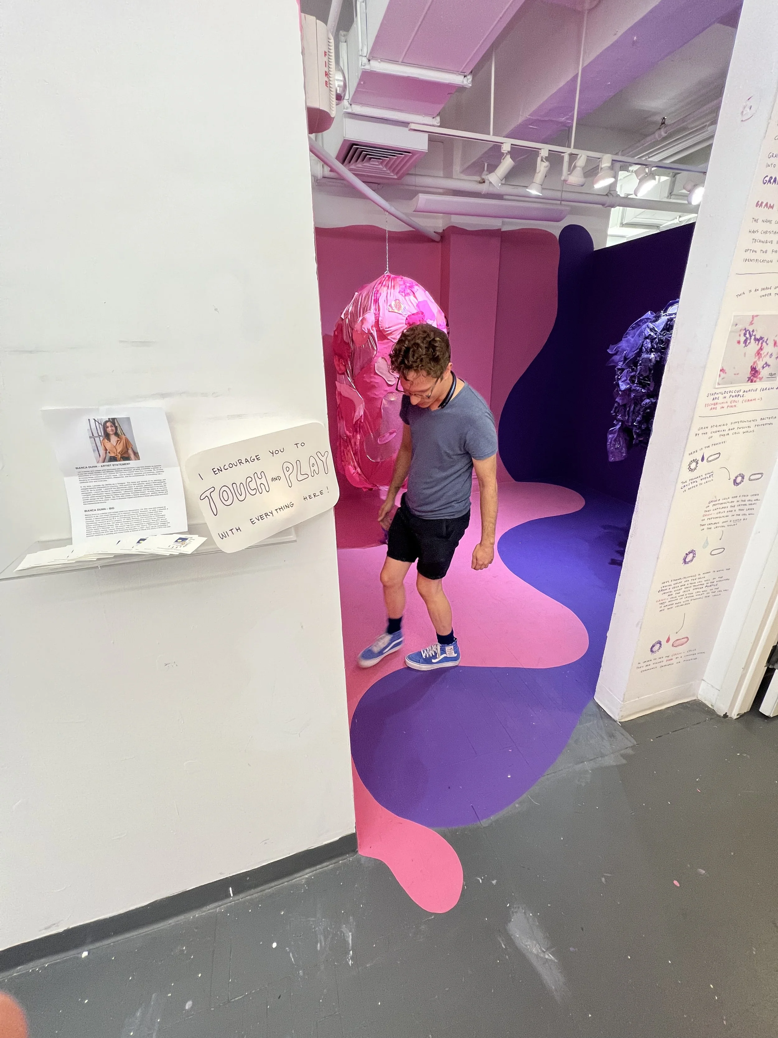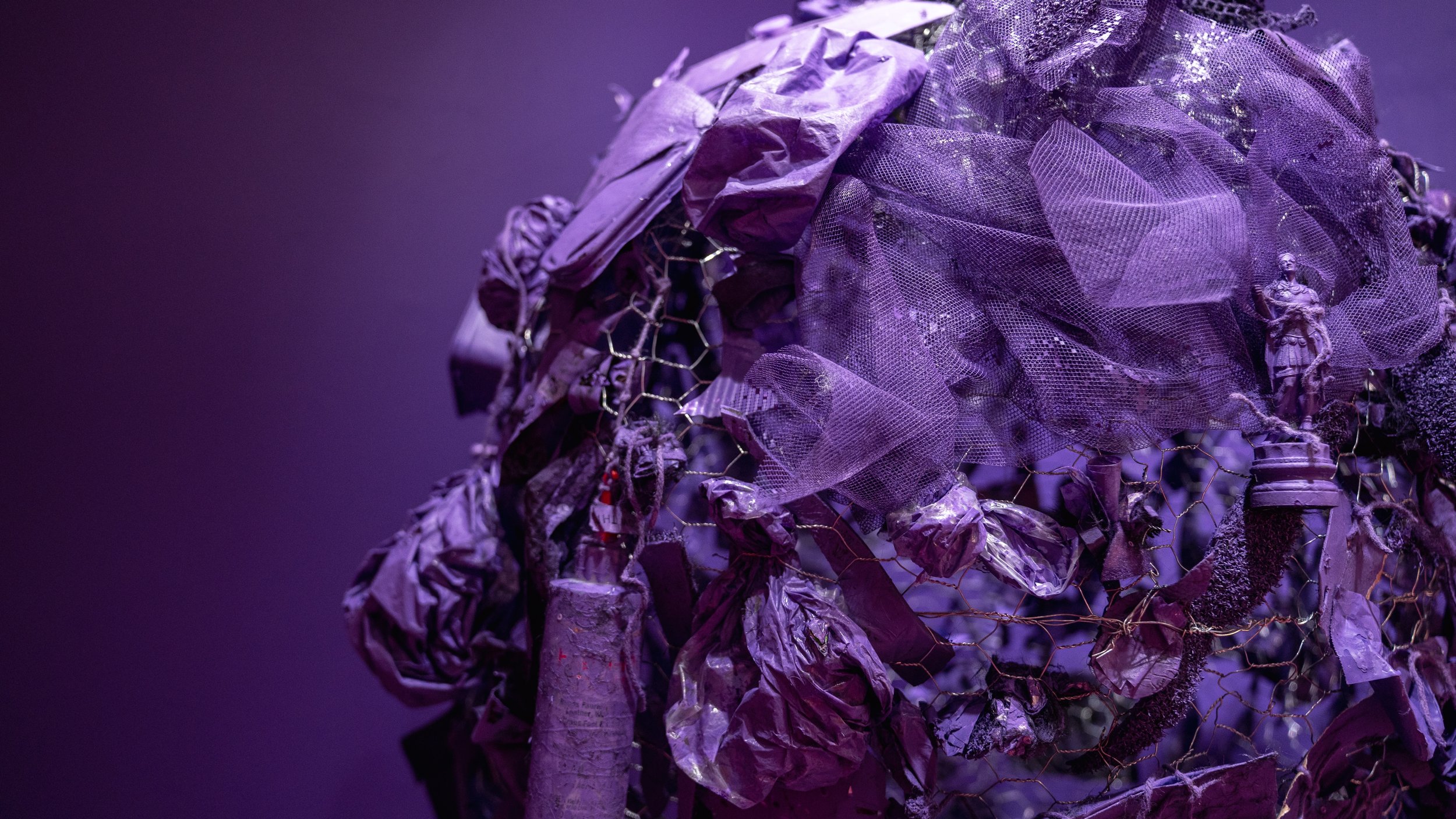
the bacteria room
I ♡ science. Bacteria are cool and complex. I hope this installation inspires you to learn more about science and to feel a sense of wonder in the world around us.

Originally shown at SVA Fine Arts Residency, Contemporary Practices, July 5–Aug 5, 2022. Photos by Victoria Lau and Jingyao Huang.
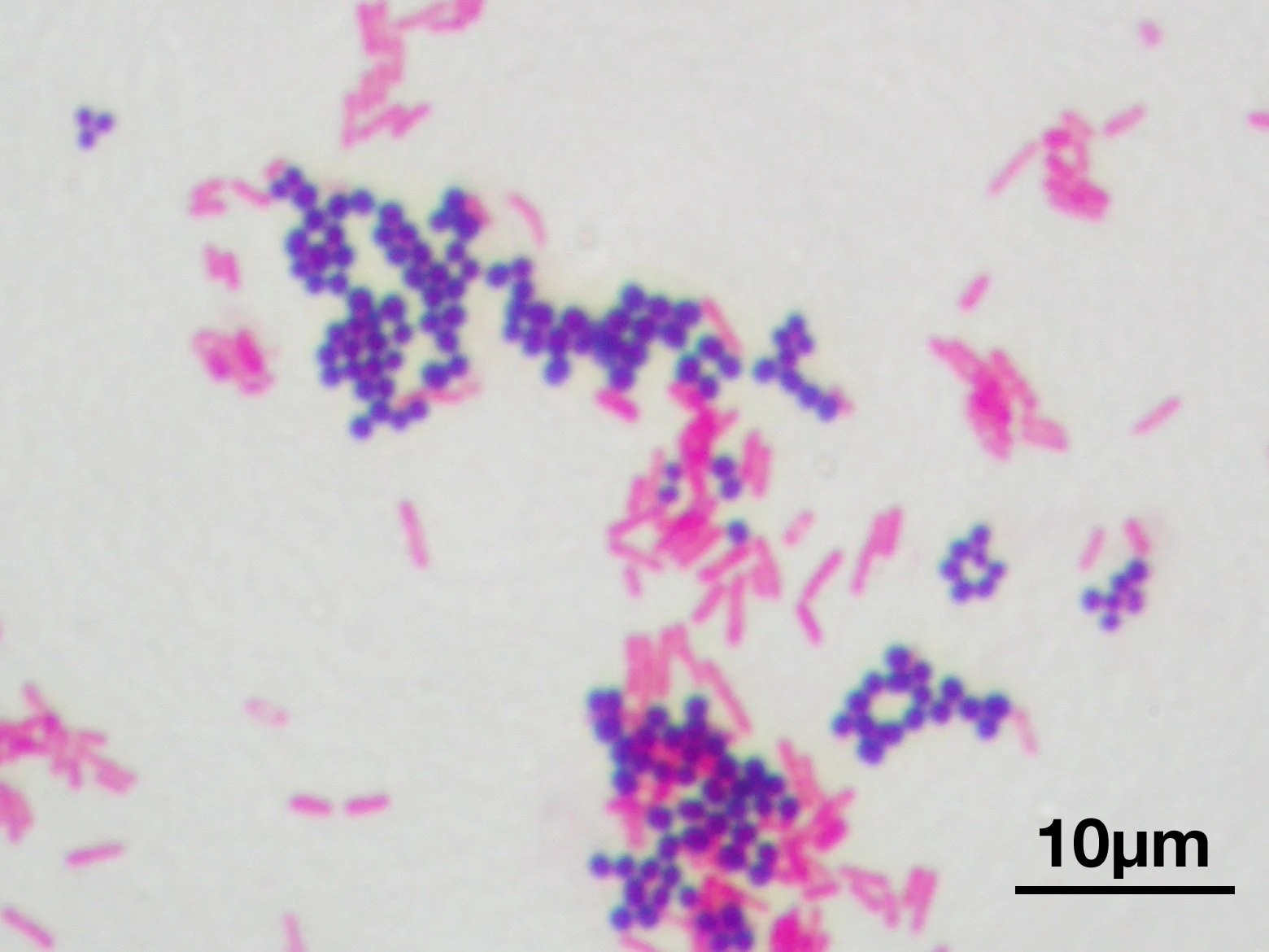
This room is based on a bacterial staining method called the Gram stain. Here is an image of a real Gram stain under the microscope:
S. aureus, a type of Gram positive bacteria, are in purple.
E. coli, a type of Gram negative bacteria, are in pink.
-
This pink and purple room is inspired by a method of staining bacteria called the Gram Stain. Gram staining classifies bacterial species into two large groups: Gram positive bacteria and Gram negative bacteria. The name comes from the Danish bacteriologist Hans Christian Gram, who developed the technique in 1884. Gram staining is often the first step in the preliminary identification of a bacterial organism.
Gram staining differentiates bacteria by the chemical and physical properties of their cell walls. Here is the process:
1. The primary stain, crystal violet is added to cells.
2. Gram+ cells have a thick layer of peptidoglycan in the cell wall that captures the crystal violet. Gram- cells have a thin layer of peptidoglycan in the cell wall that captures only a little bit of the crystal violet.
3. Next, ethanol (alcohol) is added to wash the crystal violet off the cells. Gram+ cells have a thick cell wall so the crystal violet stays trapped in the structure and the cells appear purple. Gram- cells have a thin cell wall so the small amount of crystal violet on the cell wall is washed away with ethanol; the cells are now colorless.
4. In order to see the Gram- cells, they are stained pink by a counter stain, commonly safranin or fuchsine.